Pelvic Anatomy Cat - Cat Anatomy Wikipedia / Ranzcr anatomy paper 1 march 2018 by dr kieran kusel labelling by dr amaran parasuramar;. The pelvis is firmly attached to the spine (sacroiliac joint) and the limb is longer and more angulated than the thoracic limb (which is designed to bear weight and absorb impact). Mri and ct have proven equally sensitive to the presence of disease, with a better visualiz … In newborns, the pelvis, which is narrow, cannot support the child.as an infant begins to walk, their pelvis widens. The body of the prostate is a landmark for dividing the pelvic urethra into preprostatic, prostatic, and postprostatic divisions. Anatomy of the cat (pelvic limb) origin of sartorius.
The cat is more flexible than the dog; We would love to hear your thoughts and opinions. Abdominal scans can be used to help a doctor pinpoint the location of a tumor before a biopsy is. Thirty nine patients with abnormal pelvis were compared with ct and ultrasounds. The slice thickness is 2.5 mm.
Feline Pelvis Model 9160 For Sale Anatomy Now from www.anatomynow.com An abdominal or pelvic ct scan can be used to discover a hernia, masses, tumors, infections, or injuries. This is a ct of the abdomen and pelvis, enterography protocol. We would love to hear your thoughts and opinions. The innominate bones articulate with each other anteriorly and with the sacrum posteriorly. Anatomy of the abdomen and male pelvis using cross sectional imaging ct interactive atlas of human anatomy we have created an anatomical atlas of abdominal and pelvic ct which is an interactive tool for studying the conventional anatomy of the normal structures based on a multidetector. A pelvic ct scan takes pictures of your pelvis (the area between your hips). Anatomy ct axial abdomen and pelvis male. Feline_pelvic_anatomy 1/2 feline pelvic anatomy download feline pelvic anatomy feline pelvic anatomy thank you for downloading feline pelvic anatomy.
Each picture, also called a slice, shows a few layers of your.
The video covers the most clinic. A pelvic ct scan takes pictures of your pelvis (the area between your hips). Anatomy by dr muhammad bin zulfiqar; Each picture, also called a slice, shows a few layers of your. Anatomical structures of the abdomen. The digestive system ( cat) ( dog) includes the mouth, teeth, salivary glands, esophagus, stomach, intestine, pancreas, liver and gall bladder. Thoracic spine in full body ct scan. And anatomically, the cat lacks ligaments found in the dog and other common domestic mammals. Thirty nine patients with abnormal pelvis were compared with ct and ultrasounds. Ct mri radiographs anatomic diagrams and nuclear images. Anatomy of the lower urinary tract • anatomically,. This is the third article in our imaging essentials series—a series focused on providing critical information on radiography of the dog and cat. The pelvis supports the muscles that balance and move the trunk, hips, and legs (2).
The digestive system absorbs and digests food and eliminates solid wastes from the body. The purpose of this article is to review the three basic. Anatomy by dr muhammad bin zulfiqar; This is the third article in our imaging essentials series—a series focused on providing critical information on radiography of the dog and cat. A ct scan of this area may be done to look for abscesses, tumors, kidney stones, infections, or the cause of unexplained abdominal pain.
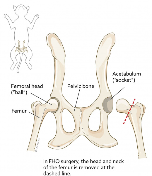
Femoral Head Ostectomy Fho In Cats Vca Animal Hospital from vcahospitals.com This is the third article in our imaging essentials series—a series focused on providing critical information on radiography of the dog and cat. The slice thickness is 2.5 mm. The digestive system ( cat) ( dog) includes the mouth, teeth, salivary glands, esophagus, stomach, intestine, pancreas, liver and gall bladder. Radiographers suggest an abdominal ct scan to look for the following: The integumentary system is the skin and fur that cover the animal's body. Thirty nine patients with abnormal pelvis were compared with ct and ultrasounds. The body of the prostate is a landmark for dividing the pelvic urethra into preprostatic, prostatic, and postprostatic divisions. Learn cat anatomy pelvic with free interactive flashcards.
It is performed with a higher radiation dose and larger dose of iv contrast, which helps to evaluate subtle areas of bowel inflammation.
The skin protects the underlying organs. The body of the prostate is a landmark for dividing the pelvic urethra into preprostatic, prostatic, and postprostatic divisions. An abdominal or pelvic ct scan can be used to discover a hernia, masses, tumors, infections, or injuries. The pelvis is firmly attached to the spine (sacroiliac joint) and the limb is longer and more angulated than the thoracic limb (which is designed to bear weight and absorb impact). The musculoskeletal system is responsible for form, support, stability and movement. Male abdomen and pelvis ct scan form no 2: This video details comprehensive anatomy of the abdomen with coronal correlation.please visit us at www.pulseradiology.com Ranzcr anatomy paper 1 march 2018 by dr kieran kusel labelling by dr amaran parasuramar; The integumentary system is the skin and fur that cover the animal's body. The digestive system absorbs and digests food and eliminates solid wastes from the body. The purpose of this article is to review the three basic. Anatomia by dr césar reyes; This is the third article in our imaging essentials series—a series focused on providing critical information on radiography of the dog and cat.
Browse 500 sets of cat anatomy pelvic flashcards. A pelvic ct scan takes pictures of your pelvis (the area between your hips). Ct mri radiographs anatomic diagrams and nuclear images. Feline_pelvic_anatomy 1/2 feline pelvic anatomy download feline pelvic anatomy feline pelvic anatomy thank you for downloading feline pelvic anatomy. It is performed with a higher radiation dose and larger dose of iv contrast, which helps to evaluate subtle areas of bowel inflammation.
X Ray Of A Cat With A Pelvic Fracture Stock Image Image Of Hind Feet 191178991 from thumbs.dreamstime.com • a small prostate gland is positioned in the middle of the pelvic urethra of the male cat. Über 7 millionen englischsprachige bücher. The body of the prostate is a landmark for dividing the pelvic urethra into preprostatic, prostatic, and postprostatic divisions. And anatomically, the cat lacks ligaments found in the dog and other common domestic mammals. The cat has 230 bones, as opposed to 206 within the human body. The pelvic girdle (hip girdle) is formed by a single bone, the hip bone or coxal bone (coxal = hip), which serves as the attachment point for each lower limb. Each picture, also called a slice, shows a few layers of your. The cat is more flexible than the dog;
This video details comprehensive anatomy of the abdomen with coronal correlation.please visit us at www.pulseradiology.com
Feline_pelvic_anatomy 1/2 feline pelvic anatomy download feline pelvic anatomy feline pelvic anatomy thank you for downloading feline pelvic anatomy. The pelvic girdle (hip girdle) is formed by a single bone, the hip bone or coxal bone (coxal = hip), which serves as the attachment point for each lower limb. 26 july 2016 male abdomen and pelvis ct scan form no 1: The integumentary system is the skin and fur that cover the animal's body. Anatomy of the cat (pelvic limb) origin of sartorius. And anatomically, the cat lacks ligaments found in the dog and other common domestic mammals. Anatomy ct axial abdomen and pelvis male male abdomen and pelvis ct scan form no 1. Learn cat anatomy pelvic with free interactive flashcards. The musculoskeletal system is responsible for form, support, stability and movement. A pelvic ct scan takes pictures of your pelvis (the area between your hips). Abdomenal ct showing the pelvis. The slice thickness is 2.5 mm. Anatomy ct axial abdomen and pelvis male.
Male abdomen and pelvis ct scan form no 2: pelvic anatomy. Ct mri radiographs anatomic diagrams and nuclear images.


0 Komentar